


Methods
- Structural Magnetic Resonance Imaging (sMRI)
- Functional Magnetic Resonance Imaging (fMRI)
- Diffusion Weighted/Tensor Imaging (DWI/DTI)
- Arterial Spin Labeling (ASL)
- Spectroscopy
- Transcranial Direct Current Stimulation (tDCS)
- Transcranial Magnetic Stimulation (TMS)
- Electroencephalogram / Evoked Response Potential (EEG/ERP)
- Psychophysical Methods
Structural Magnetic Resonance Imaging (sMRI)
For Structural MRI analysis we use a technique which is called voxel-based morphometry. VBM pre-processing is composed of three steps: spatial normalization of MRI scans into a stereotaxic reference space using a custom-made whole-brain template, segmentation of the normalized images into grey matter, white matter and cerebrospinal fluid, and smoothing of the data typically with a 12mm Gaussian kernel. Customized whole-brain as well as gray matter, white matter, and CSF templates will be created from the group of subjects enrolled in this study. To facilitate optimal segmentation, we will estimate normalization parameters while removing non-brain voxels (skull, sinus) using an optimized protocol (Good et al., 2001).
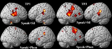
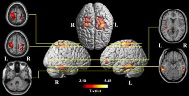
Functional Magnetic Resonance Imaging (fMRI)
Functional magnetic resonance imaging allows scientists to take pictures of brain activity. Unlike standard MRI scans, which only show the structure or anatomy of the brain, fMRI actually shows the areas which are active while performing a specific task. fMRI also allows to examine the differences in brain function or activity caused by certain brain disorders.
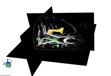
Diffusion Weighted/Tensor Imaging (DWI/DTI)
Diffusion tensor imaging (DTI) provides information regarding the structural integrity of white matter pathways in the brain by measuring the molecular diffusion of water in brain tissue. Diffussion is influenced by myelin density, the number of myelinated fibers, and axonal membrane integrity. Thus, DTI is an indirect measure of the structural integrity of white matter and is sensitive to alterations in tissue properties that conventional structural magnetic resonance imaging does not detect.
Arterial Spin Labelling (ASL)
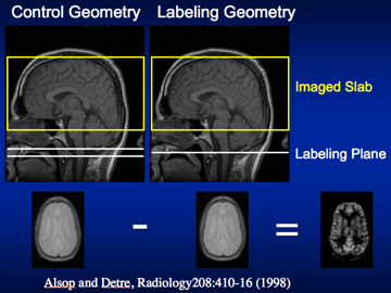
Arterial spin labeling is a non-invasive MRI method to measure cerebral blood flow. It works by magnetically tagging the inflowing blood and subtracting the resulting image from an image that has no magnetic tag. Through some computations, one can calculate absolute cerebral blood flow. Our collaborator in the Radiology Department at BIDMC, Dr. Alsop, is one of the developers of this technique. We are using ASL mainly to examine blood flow changes in relation to stimulation of the brain as well as in stroke patients that may have cerebral perfusion issues.
Spectroscopy
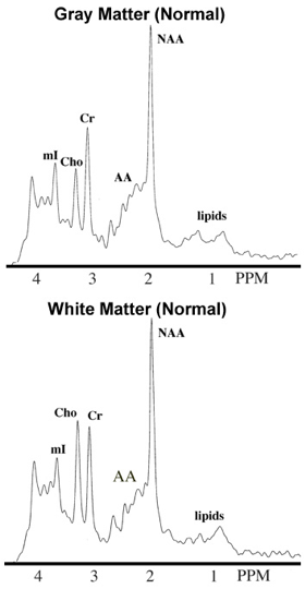
Magnetic resonance spectroscopy (MRS) detects electromagnetic signals produced by the interaction between the atomic nuclei within molecules and the static magnetic field. These signals are subjected to further processing resulting in an MR spectrum (see pictures on the left) that can be obtained from defined volumes of brain tissue. The MR spectrum is a two-dimensional plot with frequency of the chemical compound on the horizontal axis and intensity of the resonance interaction (i.e., signal) on the vertical axis. These spectral can be used to obtain concentration measures for certain chemicals in living tissue and their alterations in neurological disease/disorders or after particular interventions.
Transcranial Direct Current Stimulation (tDCS)
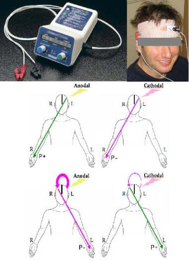
Transcranial Direct Current Stimulation (tDCS) is a noninvasive brain stimulation technique that utilizes low amplitude direct currents applied via scalp electrodes to inject currents in the brain and thus modulates the level of excitability. DC stimulation has been used in various forms since the inception of modern electrophysiology at the beginning of nineteenth century. There has been a recent upsurge in interest in tDCS as a tool for neuroscience research, and as a modality for the assessment and treatment of various neurological and psychiatric disorders. tDCS has the distinct advantages of being inexpensive, easy to administer, noninvasive and painless. Recent studies support a therapeutic potential of tDCS in recovery of motor functions, expressive language functions, and in the recovery of swallowing functions.
Transcranial Magnetic Stimulation (TMS)
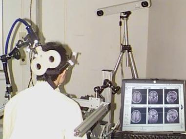
Transcranial magnetic stimulation or TMS is a neurophysiological technique that allows the induction of a current in the brain using a magnetic field to pass the scalp and the skull safely and painlessly. In TMS, a current passes through a coil of copper wirethat is encased in plastic and held over the subject's head. As the current passes through the coil it generates a magnetic field that can penetrate the subject's scalp and skull, and in turn induce a current in the subject's brain. Repetitive TMS (trains of multiple stimuli per second) can be used to study how the brain organizes different functions. In other words, it seems possible to make a given brain area work more or less for a period of minutes, or even weeks when rTMS is applied repeatedly several days in a row. This has opened up the possibility of using rTMS for therapy of some illnesses in neurology, rehabilitation, and psychiatry. TMS, particularly rTMS, can have adverse effects, the most worrisome of which is its potential to induce a seizure or epileptic convulsion, even in subjects without any predisposing illness.
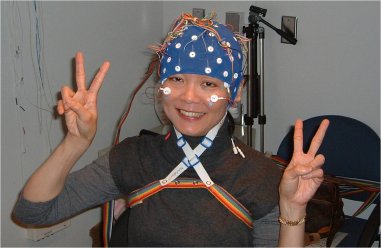
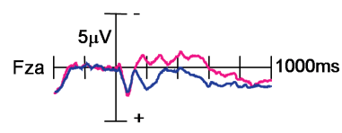
Electroencephalography / Event-Related Potential (EEG/ERP)
Electroencephalography (EEG) is a non-invasive technique for recording electrical activity from the brain. From EEG we derive Event-Related Potentials (ERPs), which are rapid fluctuations in electrical voltage specifically related to sensory or cognitive events. Using EEGs and ERPs we can obtain a temporally precise understanding of cognitive and neural function.
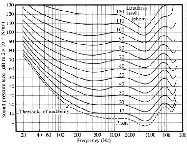
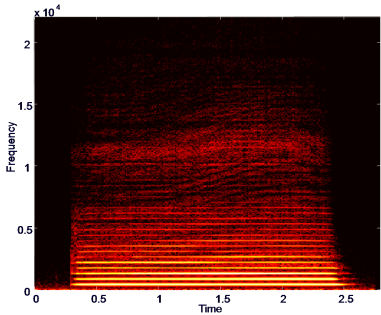
Psychophysical Methods
Psychophysics is the precise science of relating the physical aspects of the world to the psychological attributes of what a person perceives. This relationship translates into combining behavioral experiments with mathematical models, such as those used to analyze sounds and derive perceptual thresholds, for normal subjects as well as special populations.
